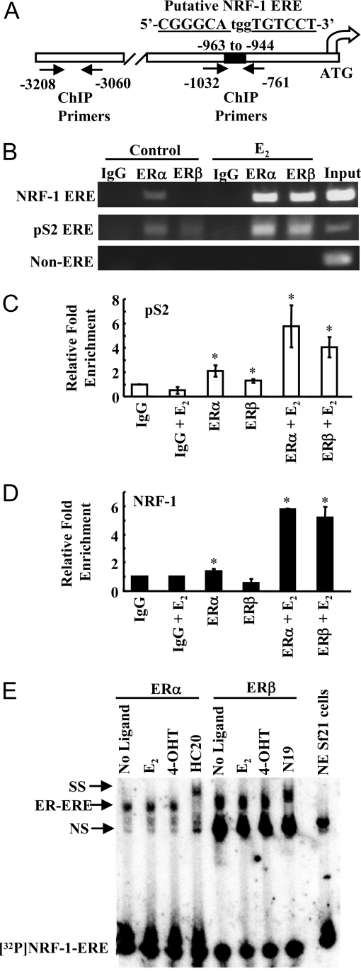
Figure 4.
ERα and ERβ Bind to the Putative NRF-1 ERE
A, Diagram of the NRF-1 promoter and putative ERE (−963 to −944) with the location of the ChIP primers. ERE half-sites are underlined and letters in bold indicate deviations from the consensus ERE. B, MCF-7 cells were treated with EtOH or E2 for 1 h, and ChIP was performed as described in MATERIALS AND METHODS. C, QRT-PCR was performed for ERα and ERβ occupancy on the pS2 ERE (C) and NRF-1 ERE (D) in ChIP samples as described in MATERIALS AND METHODS. Relative promoter enrichment compared with IgG is plotted. Values are the average and sd of two separate experiments. *, P < 0.05 for control. E, Baculovirus-expressed ERα and ERβ were incubated with 32P-labeled NRF-1 ERE in the presence of E2, 4-hydroxytamoxifen (4-OHT), or no ligand, as indicated. EMSA was performed as described in MATERIALS AND METHODS. NS, Nonspecific binding; SS, supershift with the indicated ER antibodies.

