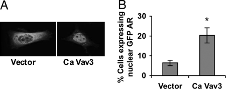
Figure 4.
Ca Vav3 Mediates Nuclear Translocation of AR in the Absence of Androgen
A, PC3 cells grown on coverslips were transfected with GFP-AR, and either Ca Vav3 or empty vector. Cells were fixed 48 h after transfection, and GFP-AR localization was assessed by confocal immunofluorescence microscopy. Twenty percent of Ca Vav3-transfected cells vs. 8% of vector-transfected cells exhibited predominantly nuclear GFP-AR as shown in the right hand image. A representative cell transfected with empty vector exhibiting cytoplasmic localization of GFP-AR is shown on the left. B, Cells were visualized and scored for GFP-AR localization. The percentage of GFP-AR expressing cells that exhibited predominantly nuclear GFP-AR in each group is plotted. Data are representative of three independent experiments, and significance was determined using a two-tailed Student’s t test (*, P < 0.05).

