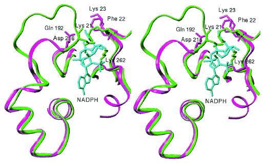
Figure 5.
Stereo view of the details of the structure of 2,5-DKGR A and aldose reductase in the vicinity of the NADPH cofactor binding site. In aldose reductase (green), the loop 7 region completely covers the NADPH cofactor (cyan) by means of electrostatic interactions between residues Lys-21, Asp-216, and Lys-262. In 2,5-DKGR A (magenta), however, this loop is shorter and does not clamp over the cofactor. In aldose reductase a major rearrangement of the loop 7 region appears necessary to allow the NADPH cofactor to enter and exit the binding cleft.

