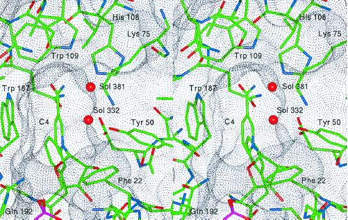
Figure 6.
Stereo view of the active site of 2,5-DKGR A in the region of the binding site for the 2,5-DKG substrate. The van der Waals dot surface indicates the general shape of the substrate-binding pocket. The residues that define the pocket are indicated, as well as two solvent molecules (Sol 332 and 381) that reside within the pocket. These water molecules are postulated to be displaced by specific substrate functional groups (see text).

