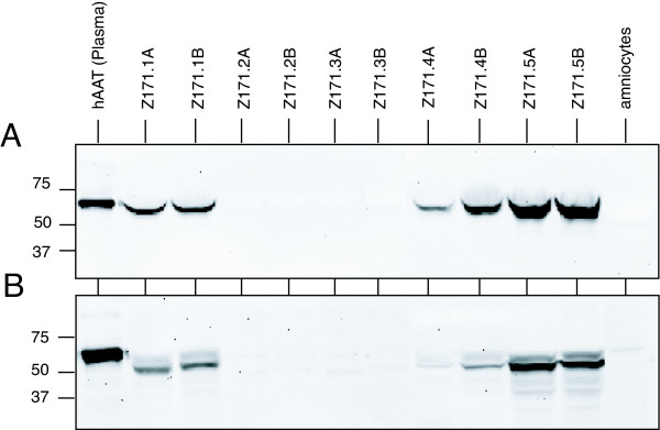
Figure 2.
Expression of hAAT protein. Western Blot analyses for hAAT protein expressed in 10 E1-transformed amniocyte cell pools. (A) For detection of secreted hAAT, proteins in the cell culture medium were fractionated on a SDS-containing polycrylamide gel. (B) For detection of intracellular hAAT, cells were lysed and proteins were separated on a SDS-containing polyacrylamide gel. Intracellular and secreted hAAT was visualized using a monoclonal anti-hAAT antibody. For control proteins from untransformed amniocytes and hAAT purified from human plasma were used.

