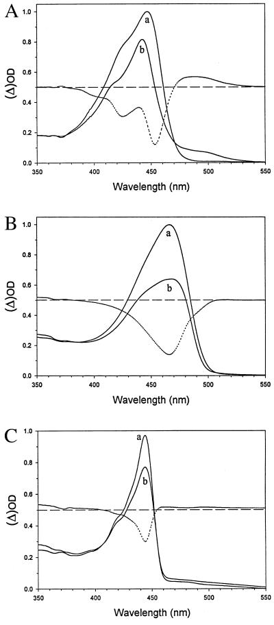
Figure 3.
Light-induced formation of pR and a pR-like intermediate in PYP (A), hybrid I (B), and hybrid II (C), at low temperature (77 K). Each protein was dissolved, at a concentration of 20 μM, in a buffer of 10 mM Tris⋅HCl, pH 7.0, containing 50% (vol/vol) glycerol. Samples were frozen in 1-cm acrylic cuvettes in the dark. After recording of the dark spectra (a traces), each sample was illuminated in the cryostat, as described in Materials and Methods, for 20 min, to induce pR formation (b traces). The absorbance in both spectra was set to zero at 600 nm for background subtraction. The dotted line in each panel represents the difference spectrum between traces a and b.

