
Fig. 4.
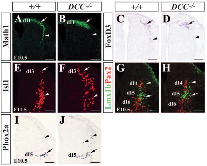
Expression of early-born neuron-specific markers in wild-type and Dcc−/− mutants. (A,B) Math1 immunocytochemical staining of E10.5 wild-type (A) and Dcc−/− mutant (B) dorsal neural tubes shows an absence of Math1+ cells in more ventral aspect of the neural tube of Dcc−/− mutant (arrowhead in B) when compared with wild-type control (arrowhead in A). (C,D) Foxd3 (dI2 marker) expression in E10.5 wild-type (C) and Dcc−/− mutant (D) neural tubes detected by in situ hybridization. Foxd3+ cells are absent in more ventral aspect of the neural tube, as indicated by arrowhead in D. (E,F) Isl1 staining of E11.5 wild-type (E) and Dcc−/− mutant (F) neural tubes. More Isl1+ cells are present in the dorsal part of the neural tube (arrow in F). (G,H) Double staining of Lmx1b (green; dI5 marker) and Pax2 (red; dI4, dI6 marker) in E10.5 wild-type (G) and Dcc−/− mutant (H) neural tubes. Arrowhead in H indicates dorsally migrated Lmx1b+ cells. (I,J) Phox2a staining of E10.5 wild-type (I) and Dcc−/− mutant (J) neural tubes. Arrows and arrowheads in I,J indicate normal and abnormal positions of Phox2a+ neurons, respectively. Scale bars: 100 μm.