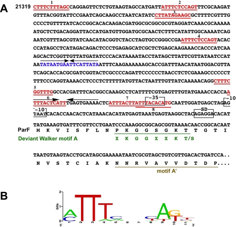
Figure 4. Genetic structures located upstream of parF and parG.
A. The direct repeats within the pMET1 putative parH-like locus are shown in red. The diagram also shows the −35 and −10 sequences, as well as the inverted repeats (arrows). The inverted repeat within the putative parH locus is shown in blue. The beginning of the ParF amino acid sequence including the deviant Walker motif A and motif A' are shown. B. Logo plot [60], [61] of a multiple alignment of the direct repeats shown in red.

