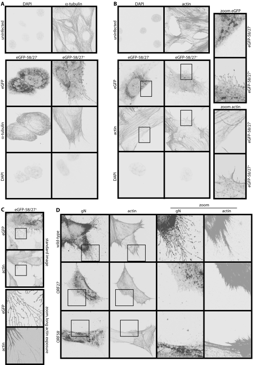
Figure 5. MHV-68-induced membrane fronds contain actin but not tubulin.
A. NIH-3T3 cells were left uninfected or infected (1 p.f.u./cell, 16 h) with either ORF27− or ORF27+ eGFP-ORF58 MHV-68. They were then fixed, permeabilized and stained for α-tubulin. EGFP fluorescence was visualized directly. Nuclei were counterstained with DAPI. B. NIH-3T3 cells were infected or not as in A, then fixed, permeabilized and stained for actin with Alexa568-conjugated phalloidin. EGFP fluorescence was visualized directly and nuclei were counterstained with DAPI. The zoomed images correspond to the boxed regions. They show coincident membrane fronds and actin spikes with ORF27+ infection and neither with ORF27− infection. C. NIH-3T3 cells were infected (1 p.f.u./cell, 16 h) with ORF27+ eGFP-ORF58-tagged MHV-68 and stained for actin as in B, but with longer exposure times for the zoomed images (which correspond to the boxed regions above) to show the very fine actin cores of the distal membrane fronds. D. NIH-3T3 cells were infected (1 p.f.u./cell, 16 h) with untagged wild-type, ORF27− or ORF58− MHV-68. The cells were then fixed, permeabilized and stained for gN with mAb 3F7 plus Alexa488-conjugated goat anti-mouse IgG pAb, and for actin with Alexa568-conjugated phalloidin.

