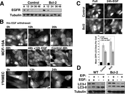
Figure 5.
EGF Withdrawal induces autophagy. (A) Lysates from pBABE control and Bcl-2–expressing MCF-10A cells grown attached (A) or suspended for the indicated times were subject to immunoblotting with α-EGFR and α-tubulin. (B) GFP-LC3–expressing MCF-10A cells or 1°HMECs were grown in media lacking EGF for the indicated times. Twelve hours of EGF readdition reverses LC3 puncta formation. (C) GFP-LC3 puncta in pBABE control and Bcl-2–expressing cells after 24 h of EGF withdrawal. Bar, 20 μm. Bottom, quantification of GFP-LC3 dots per cell in control and Bcl-2 cells grown in complete growth media (black), HBSS starved for 5 h (light gray), or EGF withdrawn for 24 h (dark gray). Results are the mean ± SEM enumerated from 40 to 60 individual cells using MetaMorph (GE Healthcare) software. (D) Wild-type and Bcl-2–expressing cells were grown in complete media or media lacking EGF for 24 h, lysed, and subject to immunoblotting with α-LC3 and α-tubulin.

