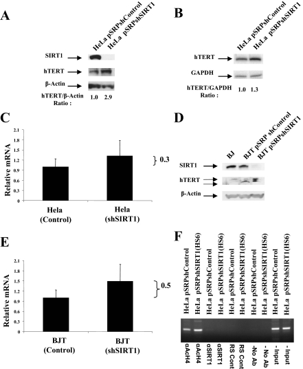
Figure 2.
Regulation of hTERT. (A) Effect of SIRT1 suppression on hTERT protein in Hela cells. Western blot analysis was performed on cell lysates, and they were subjected to immunoblotting with anti-SIRT1, anti-hTERT, and anti-β-actin antibodies. (B) Effect of SIRT1 suppression on hTERT mRNA in Hela cells. Total RNA was isolated from Hela-pSRP-shControl and Hela-pSRPshSIRT1 cells and hTERT mRNA was quantified by quantitative radioactive in-gel PCR as described (Nakamura et al., 1997). The ratio of hTERT/GAPDH is shown. (C) Quantification of hTERT mRNA in Hela and Hela-pSRPshSIRT1 cells by real-time Q-PCR. The values shown are normalized to an internal GAPDH control. (D) Regulation of hTERT protein in BJT cells. BJT-pSRP-shControl and BJT-pSRPshSIRT1 cell lysates were resolved on 4–12% gradient gels, and immunoblotting was performed using an anti-hTERT rabbit antibody. Primary BJ cells in the first lane were used as negative control. (E) Effect of SIRT1 suppression on hTERT mRNA in BJT cells. Same as in C except that BJT and BJT-pSRPshSIRT1 cells were used. (F) ChIP of hTERT promoter using the antibodies shown. Hela control and SIRT1(HS6) knockdown cells were used in each ChIP reaction as shown. Antibodies used were against total acetylated H4 and SIRT1. Controls were rabbit serum (RS) and no antibody reactions. For details consult Materials and Methods.

