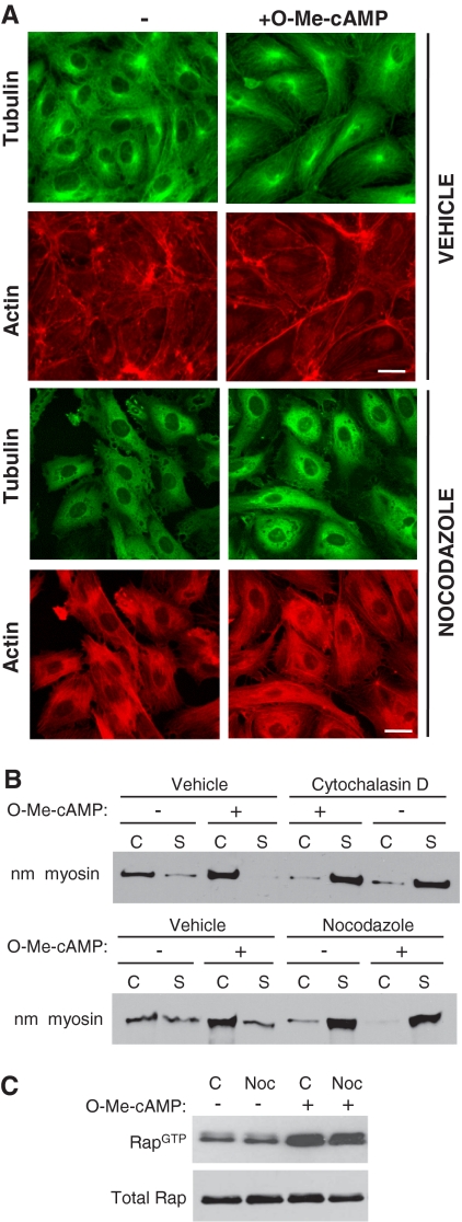
Figure 5.
Analysis of O-Me-cAMP–induced F-actin and Rap1 activation after nocodazole treatment of HUVECs. (A) HUVEC were treated with vehicle alone or with 0.15 μM nocodazole for 5 min and simultaneously treated with control (−) or O-Me-cAMP. Cells were fixed and stained with fluorescein isothiocyanate-labeled anti-tubulin antibody (microtubules) and rhodamine-phalloidin (F-actin). Nocodazole treatment prevented O-Me-cAMP induced F-actin changes detected in control/O-Me-cAMP–treated cells. One of five representative experiments is shown. Bar, 10 μm. (B) The Triton-X cytosolic soluble (S) and cytoskeletal (C) fractions were prepared from endothelial cells treated with O-Me-cAMP, cytochalasin D, and nocodazole as indicated, and then they were immunoblotted with nonmuscle myosin heavy chain (nm myosin) antibody. (C) Pull-down assay of active Rap were performed on HUVECs incubated without (−) or with 0.15 μM nocodazole (+) for 5 min followed by a 20 min treatment with O-Me-cAMP (+) as indicated. Western blots were probed for Rap1. One of three independent experiments is shown for B and one of two for C.

