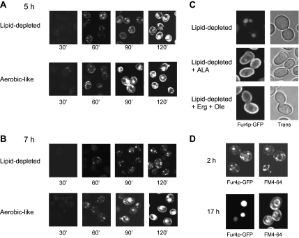
Figure 2.
Impact of lipid depletion on Fur4p-GFP delivery to the cell surface. hem1Δ [pFl38gF-GFP] cells were grown to late exponential phase in YPRaff + ALA and shifted to fresh YPRaff (lipid-depleted) or YPRaff + ALA (aerobic-like). Fur4p-GFP synthesis was induced by adding galactose (4% final) to the medium after 5 h (A) or 7 h (B) after the shift. The GFP signal was visualized by confocal microscopy at the time indicated after galactose addition. (C) hem1Δ end3Δ [pFl38gF-GFP] cells were grown for 7 h under lipid-depleted conditions (YPRaff) before induction of Fur4p-GFP synthesis by galactose addition. After 3 h after Fur4p-GFP induction (i.e., after 10 h growth under lipid-depleted conditions), ALA (lipid-depleted + ALA), or ergosterol and Tween 80 (lipid-depleted + Erg + Ole) were added to the medium. GFP fluorescence was observed by confocal microscopy 24 h after shift to lipid-depleted conditions. The exact same settings were used throughout the experiment to obtain a semiquantitative signal. (D) hem1Δ [pFl38gF-GFP] cells grown for 7 h under lipid-depleted conditions as described in C. The GFP signal and FM4-64 cellular distribution were observed as described in Materials and Methods after 2 or 17 h after galactose addition. Trans, transmission.

