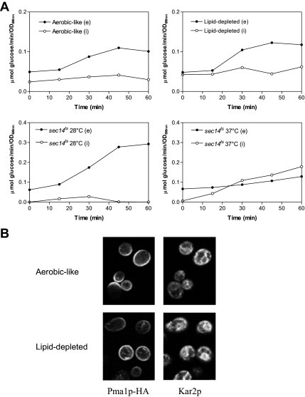
Figure 4.
Consequences of lipid depletion on invertase secretion and Pma1p plasma membrane targeting. (A) hem1Δ cells (top) were grown as described in the legend of Figure 1 for 7 h in YPD +ALA (aerobic-like) or YPD (lipid-depleted) before invertase induction. sec14ts cells (bottom) were grown to exponential phase at 28°C and invertase was induced by glucose limitation in cells maintained under permissive conditions (28°C) or transferred to nonpermissive temperature (37°C). Samples were removed at the time points indicated and the activity of internal and secreted invertase was determined as described in Materials and Methods. Closed symbols, extracellular invertase activities (e); open symbols, intracellular invertase activities (i). (B) hem1Δ [Yiplac204–2HSEpr-HA-PMA1] cells were grown under aerobic-like or lipid-depleted conditions for 7 h before incubation for 15 min at 39°C for induction of Pma1p-HA synthesis. Sixty minutes after the heat shock, the cells were fixed and processed for immunofluorescence with antibodies to HA (left). Anti-Kar2p antibodies were used as nonspecific markers to visualize the cells in the microscope field (right).

