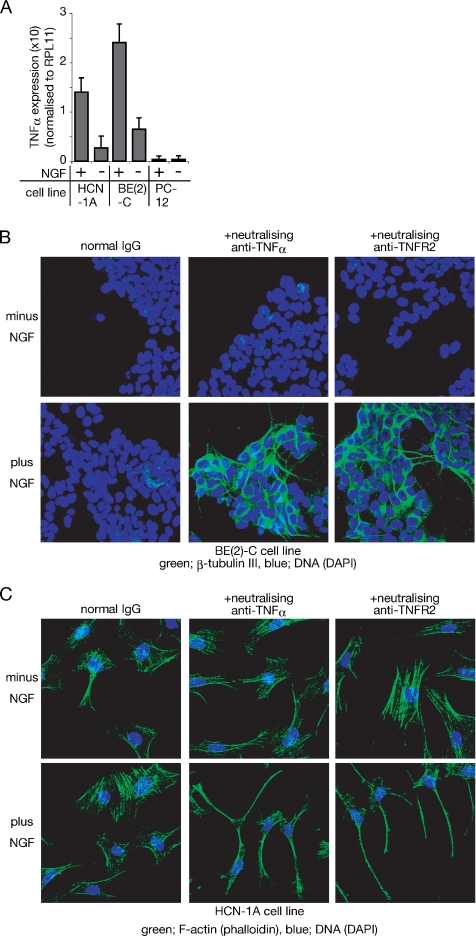
Figure 3.
HCN-1A and BE(2)-C, but not PC-12 cells, express TNF-α in response to NGF, thus inhibiting neural differentiation of these cells. (A) Semiquantitative analysis of TNF-α cDNA prepared from cells incubated with or without NGF for 24 h. Expression level of TNF-α is shown relative to RPL11 expression. The data represent the mean of three experiments. Error bars indicate SE; p < 0.001 in both BE(2)-C and HCN-1A and p > 0.05 in PC-12. (B) β-Tubulin III expression of BE(2)-C cells after incubation with the indicated reagents for 48 h. BE(2)-C cells cultured on collagen-coated dishes were incubated with the indicated reagents for 48 h, and expression of β-tubulin III was detected by anti-β-tubulin III antibody. Binding of the antibody was visualized with FITC-labeled secondary antibody. (C) Phalloidin staining of HCN-1A cells after incubation with the indicated reagents for 48 h. Cells were treated with or without NGF in the presence of either rabbit normal IgG (control), anti-TNF-α, or anti-TNFR2 for 48 h. Fixed cells were incubated with Alexa Fluor 488-labeled phalloidin.

