Figure 5.
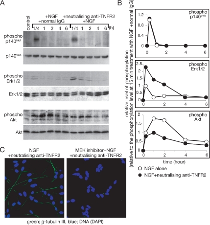
Effect of TNFR2 signaling on NGF signaling. (A) Estimation of NGF-induced phosphorylation of p140trkA, Erk1/2, and Akt with immunoblotting. SH-SY5Y cells were cultured with NGF alone or NGF and the neutralizing antibody against TNFR2. At the indicated time points after addition of NGF, cells were harvested, and whole cell extracts were prepared. The levels of phosphorylated proteins and total proteins were detected by the indicated antibodies. (B) The intensity of the signals of phosphorylated proteins indicated in A was measured with the application ImageJ (version 1.32j, National Institutes of Health, Bethesda, MD; http://rsb.info.nih.gov/ij/). Phosphorylation levels of each time point were shown relative to the phosphorylation level at 15-min treatment with NGF alone. Experiments were repeated three times, and the average phosphorylation level at each time point was plotted. Open circles indicate results from cells treated with NGF alone. Closed circles indicate results from cells treated with the combination of NGF, and the neutralizing antibody against TNFR2. (C) Effect of the MEK inhibitor on the induction of neural differentiation by the combination of NGF and neutralizing anti-TNFR2. SH-SY5Y cells were cultured on a chambered slide glass and incubated with the indicated reagents for 48 h. After fixation of cells, β-tubulin III expression was detected by anti-β-tubulin III antibody.

