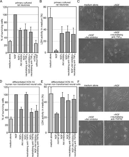
Figure 6.
TNF-α signaling is essential for NGF-dependent survival. (A and B) Cell viability assay with WST-1. The primary cultured neurons dissociated from rat dorsal root ganglia were incubated with the indicated reagents for 48 h. (A) WST-1 reagent was added directly to the culture medium after the incubation, and cells were cultured for 4 h. (B) Fifty microliters of culture medium was taken and used for LDH assay as described in Materials and Methods. (C) Cell images of neurons after incubation with the indicated reagents are taken with phase-contrast microscopy. (D and E) Differentiated HCN-1A cells were cultured in serum-free DMEM containing the indicated reagent for 24 h. (D) WST-1 reagent was added directly to the culture medium after the incubation, and cells were cultured for 2 h. (E) Fifty microliters of culture medium was treated as mentioned in B. (F) Cell images of differentiated HCN-1A cells after incubation with the indicated reagent for 24 h are taken with phase-contrast microscopy. The data represent the mean of at least three experiments. Error bars indicate SE.

