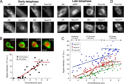
Figure 6.
Ordered reassembly of NPCs in telophase. (A) The new envelope closes in telophase (O'Donell and McLaughlin, 1984b). At this stage, Nup107 concentrates in the NE, whereas only traces of Nup214 and Pom152 are found. Note that low levels of Pom152 are present all over the ER. The nuclear marker NLS-3xRFP (nRFP) does not yet occur in the lumen of the small envelopes, indicating that protein import has not yet started. Bar, 1.5 μm. (B) As the chromosomes decondense, NLS-3xRFP occurs in the interior of the expanding nucleus, indicating that active protein import takes place. Nuclear import proceeds until cells begin to grow and form apical buds (integrated intensity; triangles), which indicates the beginning of G2 phase (Snetselaar and McCann, 1997; open symbols). Image series was taken from a single cell; the graph is based on the analysis of numerous nuclei whose circumference at a central plane was taken as an indication of their size and degree of chromosome decondensation. Elapsed time is given in minutes:seconds. Bar, 1.5 μm. (C) Quantitative analysis of the signal intensity of Nup107-GFP, Pom152-GFP, and Nup214-GFP (strains strains FB2N107G_ER, FB2N214G_ER, and FB2P152G_ER) in relation to the size of the nucleus, indicated by its circumference. An estimate of the timing and correlation to the cell cycle is presented above the graph. The amount of all nucleoporins continuously increases until cells begin to bud and reach G2 phase (open circles). (D) Colocalization of Pom152-GFP and Nup214-RFP in nuclei with a circumference of ∼4 μm shows that Pom152-GFP gives only a weak punctuate localization as indication of incorporation into NPCs, whereas the signal of Nup214-RFP is more pronounced (strain FB2P152G_214R), suggesting that Pom152 accumulates slightly before Nup214. Bar, 1 μm.

