Figure 7.
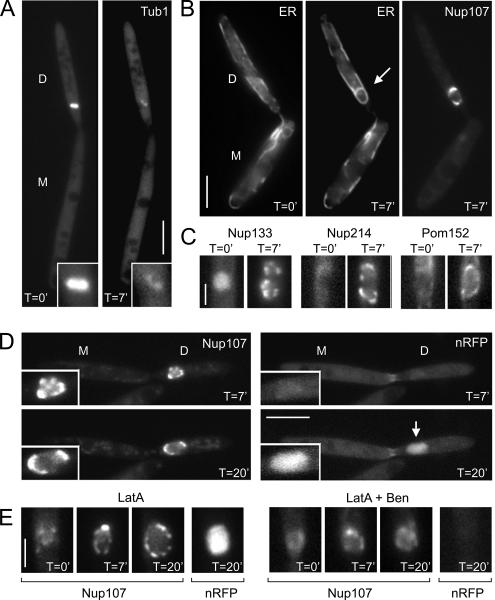
Role of the cytoskeleton in nuclear envelope reassembly. (A) Cells that express 2xRFP-Tub1 (Tub1) were placed on agarose cushions containing 30 μM benomyl. After 7 min (T = 7′), microtubules of the spindle are mainly disrupted (inset). D, daughter cell; and M, mother cell. Bar, 5 μm. (B) Anaphase cells are characterized by a cloud-like Nup107-GFP signal in the daughter cell that is divided by a cleft representing the spindle (see Figure 4). When placed on benomyl cushions (T = 0′), no ER membranes, marked by ER-RFP (ER), were surrounding the chromosomes. However, after 7-min treatment with benomyl (T = 7′), ER membranes surround the undivided chromosomes. These membranes are also decorated with a distinct Nup107-GFP signals, suggesting that they are newly formed NEs. Note that nuclear division and migration stopped in the absence of microtubules. Bar, 5 μm. (C) All nucleoporins tested reappear after 7 min of treatment with benomyl (T = 7′; strains FB2N133G_ER, FB2N214G_ER, and FB2P152G_ER). Bar, 2 μm. (D) In the absence of microtubules, chromosomes still decondese, and the Nup107-GFP containing nuclei enlarge. In agreement with the appearance of nucleoporins, protein import occurs, which is indicated by increasing levels of the NLS-3xRFP reporter protein (nRFP; T = 20′), suggesting that functional pores are formed. Time is given in minutes. Bar, 5 μm. (E) Treatment with 10 μM of the F-actin inhibitor latrunculin A did not have an obvious effect on NPC formation (Nup107) and protein import (nRFP; left, LatA), whereas simultaneous treatment with latrunculin and benomyl (right, LatA + Ben) abolished the formation of Nup107-labeled NPCs and nuclear import, suggesting that microtubules and F-actin cooperate in NE formation in U. maydis. Time is given in minutes. Bar, 2 μm.

