Figure 2.
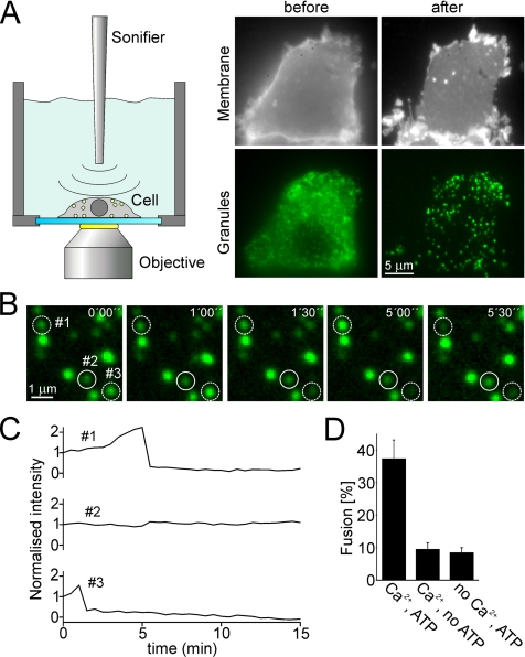
Ca2+-dependent exocytosis of secretory granules by using a membrane sheet-based cell-free assay. (A) Membrane sheets generated by sonication of PC12 cells retain docked secretory granules. Cells expressing the secretory granule marker NPY-GFP were grown on glass-coverslips and mounted on the microscope stage. GFP-labeled cells were selected and ruptured by brief pulses of ultrasound, resulting in a flat plasma membrane sheets with numerous green dots. Top, staining of the plasma membrane of a PC-12 cell before and after rupture with the lipophilic dye TMA-DPH. Bottom, GFP-channel showing secretory granules. (B) Granules docked to a membrane sheet undergo Ca2+-dependent exocytosis. Membrane sheet was preincubated for 5 min in an ATP-containing and calcium-free solution followed by the addition of ∼35 μM free calcium to trigger exocytosis (start at t = 0). Images were acquired every 30 s for 15 min. Exemplary images (time as indicated) show that the fluorescence intensity either changed (dashed circles) or remained constant (continuous circle). (C) Intensity traces of the granules encircled in B. Granules were scored as having undergone exocytosis when the drop of fluorescence intensity between two consecutive images exceeded 25%. (D) Exocytosis is dependent on the presence of Ca2+ and ATP in the triggering phase. Exocytotic membrane fusion was calculated by relating the number of granules scored positive for exocytosis during the 15 min stimulation phase to the number of granules present in the first image. Values are given as mean ± SEM (n = 9–20 membrane sheets for each condition recorded in at least three independent experiments). SEM denotes the SE of measurement.

