
Fig. 19.
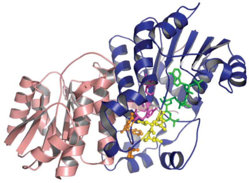
The KR domain. Crystal structure of the DEBS1 KR domain. The catalytic subdomain is shown in blue, the structural subdomain in red, the active site residues in purple, the phenylalanine, proline and glycine residues important for substrate binding in orange, the ‘LDD’ loop in yellow and NADP in green. Adapted from ref. 89.