
Fig. 20.
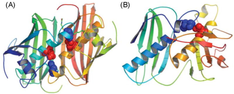
The DH domain. The E. coli DH, FabA (A) and modeled DH domain of FAS (B) exhibit two hotdog folds. The structures are colored from blue to red, from N- to C-terminus. The active site histidine (blue) and aspartate (red) residues are shown.
Official websites use .gov
A
.gov website belongs to an official
government organization in the United States.
Secure .gov websites use HTTPS
A lock (
) or https:// means you've safely
connected to the .gov website. Share sensitive
information only on official, secure websites.
The DH domain. The E. coli DH, FabA (A) and modeled DH domain of FAS (B) exhibit two hotdog folds. The structures are colored from blue to red, from N- to C-terminus. The active site histidine (blue) and aspartate (red) residues are shown.