
Fig. 9.
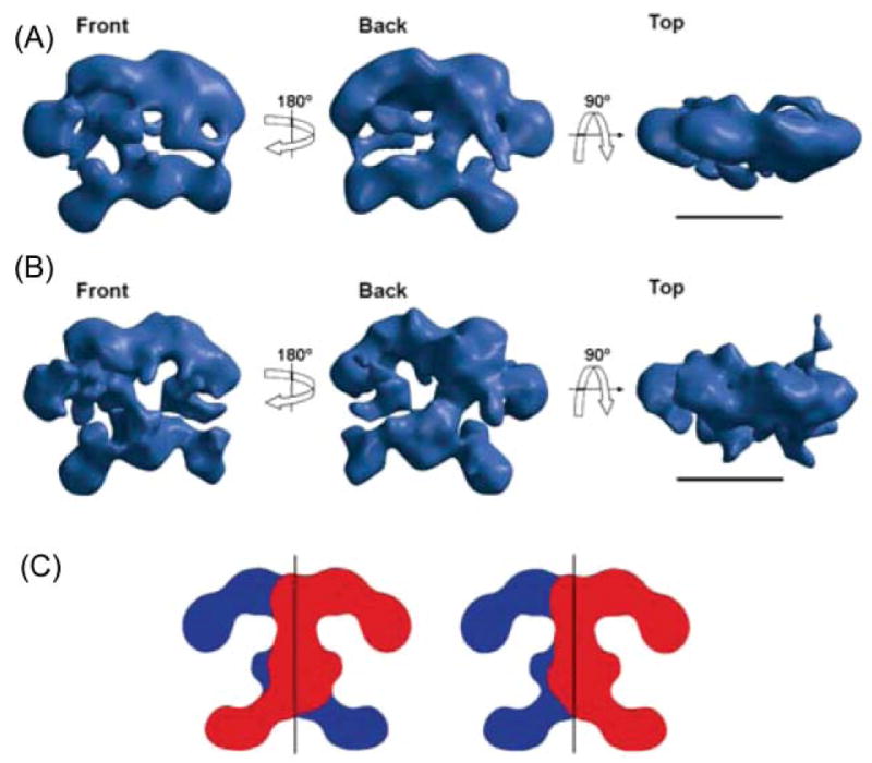
Structure of FAS determined by electron microscopy. (A) Calculated using the random conical tilt method from images of single particles of the Cys161Gln rat FAS mutant imaged under turnover conditions and preserved in negative stain; resolution ~30 Å. (B) Reconstruction of the same mutant, also imaged under turnover conditions, but preserved in amorphous ice; resolution ~16 Å. Scale bar 100 Å. (C) Alternative arrangements of the two subunits that are consistent with the structure of the dimer. Reproduced with permission from ref. 100.