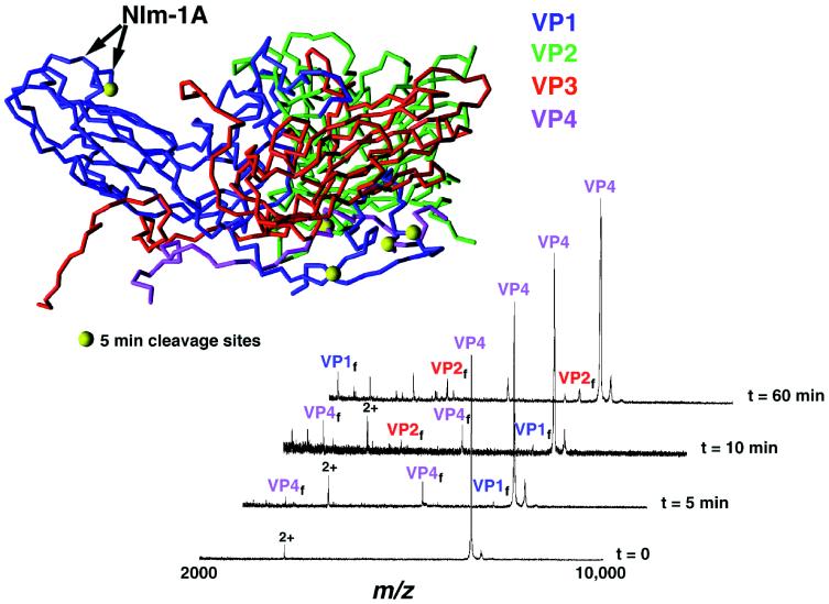
Figure 1.
Trypsin digestion time course of HRV14 (Inset). The asymmetric unit of HRV14 is shown such that the interior RNA is located at the bottom of the diagram, the nearest 5-fold axis on the left, and the nearest 2-fold axis on the right. VP1, VP2, VP3, and VP4 are blue, green, red, and mauve, respectively. Large yellow balls denote initial cleavage sites after a 5-min incubation with trypsin. Doubly charged species in the mass spectra are denoted by 2+. T = 0 represents undigested virus with VP4 observed at m/z= 7,390.0; VP1–VP3 (not shown) are observed at m/z = 32,518.9 (VP1 expected 32, 519.5 Da), 28,475.9 (VP2 expected 28, 477.4 Da), and 26,219.5 (VP3 expected 26, 217.8 Da).

