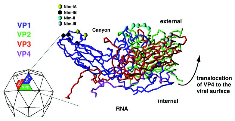
Figure 2.
X-ray crystal structure of HRV14 shows VP4 to be the most internal viral protein. Time-course mass mapping of HRV14, however, reveals proteolytic fragments originating from VP4 within the first few minutes of viral digestion, demonstrating transient exposure of VP4 to the viral surface. The large arrow illustrates the translocation of VP4 from the interior of the capsid to the external viral surface (the arrow does not indicate the mechanism of the translocation). Also shown are the position of naturally occurring escape mutation sites for NIm-IA, NIm-IB, NIm-II, and NIm-III in yellow, black, cyan, and white, respectively.

