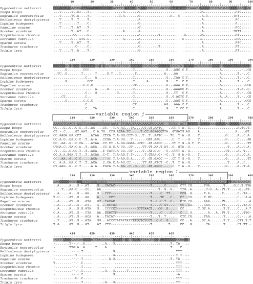
Fig. 3.
Alignment (5′ > 3′) of representative 16S rDNA sequences from the target species with binding sites (light grey) of probes (5′ > 3′; probes hybridise to the reverse complementary target strand). Double stranded (dark grey) and single stranded regions (grey) of the secondary structure are indicated in the reference sequence of Pygoplites nattereri (Ortí et al. 1996; Accession number: U33590)

