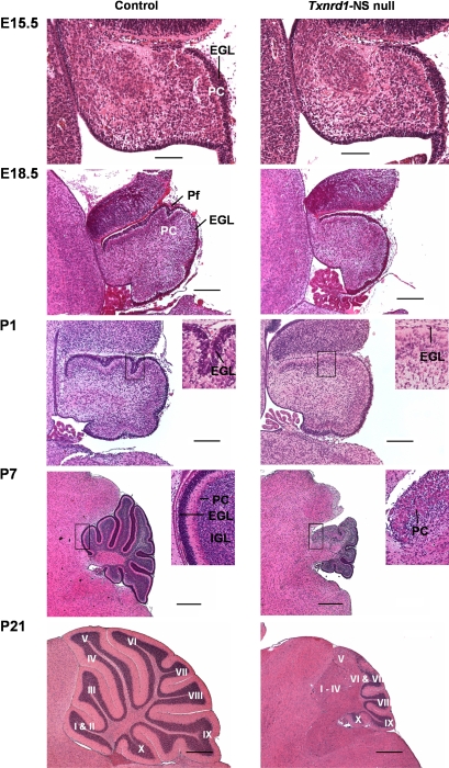
Figure 3. Striking cerebellar hypoplasia in Txnrd1-NS null mice.
H&E staining of midline sagittal cerebellar sections from control (left column) and Txnrd1-NS null mice (right column) at embryonic stages E15.5, E18.5 and at postnatal days P1, P7 and P21 showed progressive differences in the foliation and formation of the molecular-, Purkinje cell- and granular layers during development of the cerebellum. In each photograph, the anterior part of the cerebellum is located to the left and the dorsal part to the top. At E15.5, no difference in cerebellar development is visible. At E18.5, the cerebellum is smaller and the formation of the primary fissure is slightly retarded in Txnrd1 null brains. The external granular layer (EGL), which is the source of cells for the granular layer, is hypoplastic particularly in the anterior cerebellar area in Txnrd1-NS null mice at P1. Higher magnification demonstrated clear differences in the EGL thickness at P1 and P7 as well as the ectopic localisation of Purkinje cells and the absence of a well-organized, trilaminar-stuctured cerebellum. Already from E18.5 onwards, the Txnrd1 null cerebella are smaller when compared to controls, and the laminar organisation of the mutant cerebellum becomes progressively more distorted in the anterior part. At P21, the posterior lobules X to VII of the knockout mice appear relatively well developed, whereas within lobules VI–V there is an abrupt transition from an apparently normally structured cortex to a disrupted cortex. Finally, in the abnormally structured anterior region (Lobules V–I) the IGL is missing and the PCs are ectopic. In addition, the IGL is missing and the PC are ectopically localised (abbreviations: EGL = External granular layer, PC = Purkinje cells, IGL = internal granular layer, Pf = primary fissure). Lobules are indicated by roman numerals I–X. Scale bar: E15,5: 100 µm; E18,5, P1: 200 µm; P7, P21: 0,5 mm.

