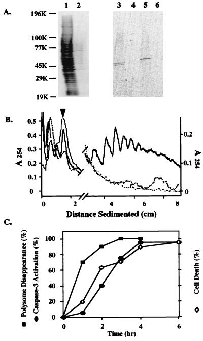
Figure 1.
Inhibition of protein synthesis during Fas-induced apoptosis in Jurkat cells. (A) Protein synthesis measured by [35S]methionine labeling. Lanes: 1, control extract; 2, apoptotic extract (4-hr anti-Fas treatment); 3, anti-cyclin A immunoprecipitate from control extract; 4, anti-cyclin A immunoprecipitate from apoptotic extract; 5, anti-cyclin B immunoprecipitate from control extract; and 6, anti-cyclin B immunoprecipitate from apoptotic extract. (B) Polysome profiles. Solid line, control extract; long- and short-dashed line, control extract treated with staphylococcal nuclease (0.05 unit/ml); dashed line, apoptotic extract (4-hr anti-Fas treatment). The arrow head indicates the position of the monosome. (C) Kinetics of polysome disappearance during anti-Fas treatment compared with those of cell death rate measured by MTT assay (20) and caspase-3 activation (measured by disappearance of procaspase-3 and appearance of PARP cleavage product). Polysome disappearance was estimated by the total area underneath the polysome peaks.

