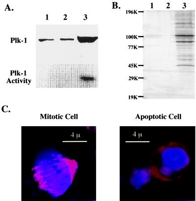
Figure 6.
Changes in mitotic parameters during Fas-induced apoptosis in Jurkat cells. Lanes: 1, control extracts; 2, apoptotic extracts (88% cell death); 3, nocodazole arrested cell extracts (≈50% in mitosis). (A) Plk-1 is not activated during apoptosis. (Upper) anti-Plk-1 Western blot. (Lower) Plk-1 activity was measured as described (14). (B) Mitotic phosphorylation as shown by MPM-2 immunoblotting (29). (C) Absence of mitotic spindle in apoptotic cells. Microtubules stained with anti-tubulin antibody appears orange-red, whereas DNA labeled with Hoechst dye (1 μg/ml) appears blue. The mitotic cell was chosen among control Jurkat cells.

