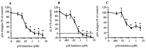
Figure 2.
Effect of p38 MAPK inhibition on IL-1β-induced p24 antigen and IL-8 in U1 cells. (A) U1 cells were cultured for 24 hr in the presence of IL-1β alone (10 ng/ml) or with IL-1β plus p38 inh (0.00098–4.0 μM). Total production of p24 antigen was measured after 24 hr, and results were expressed as percent change compared with stimulation with IL-1β (set at 100%). (B) Total IL-8 was measured in the same cultures as shown in A. The horizontal line above the x axis indicates 100% inhibition. Mean ± SEM p24 antigen or IL-8 production is shown (n = 7). •, P < 0.001 compared with control. (C) Cells were cultured in polystyrene wells for 48 hr in the presence of IL-1β alone (10 ng/ml) or IL-1β with serial 5-fold dilutions of p38 inh (0.0016–5.0 μM). Total production of p24 antigen was measured, and results were expressed as percent p24 change compared with cultures stimulated with IL-1β alone. Mean ± SEM p24 antigen is shown (n = 3). ∗∗, P < 0.01; •, P < 0.001 compared with control.

