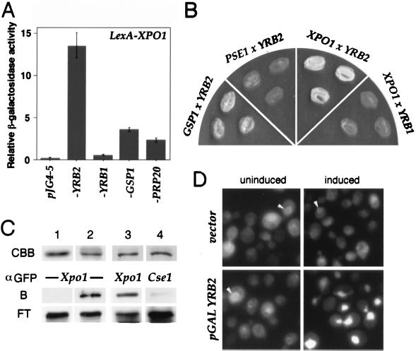
Figure 3.
Interactions between YRB2 and XPO1. (A) EGY42 × EGY48 diploid strains containing plexA-XPO1 (bait) and the indicated pJG4–5 derivatives (prey) were grown in selective media. After 2 hr of induction with galactose, β-galactosidase activity was measured as described in Materials and Methods. (B) EGY48 containing the indicated tester genes were streaked onto leucine drop-out media and grown at 30°C. (C) Cells expressing the GST-YRB2 fusion as well as XPO1-GFP or CSE1-GFP were lysed, and the resulting lysate was mixed with glutathione-Sepharose beads. After washing, proteins bound to the beads were detected by α-GFP immunoblotting. CBB, GST (lane 1) or GST-Yrb2 fusion protein (lanes 2–4) stained with Coomassie brilliant blue; B, bound; FT, flow through. (D) Cells containing a GAL1-YRB2 or a vector as well as a plasmid encoding XPO1-GFP were grown in raffinose media followed by 2 hr in galactose (induced) or glucose (uninduced) and were viewed by fluorescence microscopy. The nuclear rim is indicated with arrowheads.

