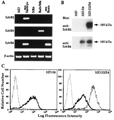
Figure 1.
Characterization of endogenous and exogenous ErbB receptor expression in 32D cells and transfectants. (A) Analysis of endogenous ErbB2, ErbB3, and ErbB4 expression in 32D cells. The amplified cDNA products generated by RT-PCR analysis of total RNA from 32D cells, day 10 murine embryo (mu Embryo), NR6 cells, Balb/MK cells, and adult murine brain (mu Brain) by oligonucleotide primers that recognize murine ErbB2, ErbB3, ErbB4, or β-actin are shown. (B) Immunoblot analysis to examine expression of ErbB2 and ErbB4 in the two 32D transfectants. Anti-ErbB2 or anti-ErbB4 serum was utilized for immunoblot (Blot) analysis of proteins from lysates of 32D, 32D.E4, or 32D.E2/E4 cells as designated. The 185-kDa mature forms of ErbB2 and ErbB4 are marked by arrows. (C) Flow cytometric analysis to determine the relative levels of cell surface expression of ErbB2 and ErbB4 on the two 32D transfectants. The histograms for untransfected 32D cells (⋅⋅) or 32D.E4 cells (—) incubated with anti-ErbB4 serum are shown (Left). The histograms for 32D (⋅⋅⋅) or 32D.E2/E4 cells incubated with anti-ErbB4 (—) or anti-ErbB2 (– – –) (Right).

