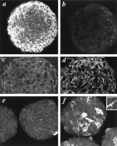
Figure 2.
Induction of Purkinje fiber phenotype in micromass cultures of embryonic myocytes. Confocal images of myocyte-micromass aggregates incubated for 48 hr in media containing no added ET (a, c, e) or 10−8 M ET (b, d, f) and immunolabeled for cMyBP-C (a, b), Cx42 (c, d), and sMHC (e, f). Inset shows the sarcomeric distribution of sMHC labeling adjacent the asterisk in a ET-treated aggregate.

