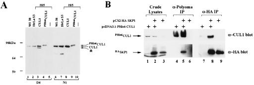
Figure 1.
hCUL1 and hSKP1 interact in vivo. (A) hCUL1 detection by affinity-purified anti-hCUL1 antibodies. Crude human cell lysates (50 μg) (lanes 1, 2, 6, and 7) and 0.5 μg of crude lysates from Hi5 insect cells, uninfected (lanes 5 and 10) or infected with hCUL1 (lanes 3 and 8) or PHis6hCUL1 (lanes 4 and 9) viruses, were resolved on an 8% SDS-polyacrylamide gel, transferred to a PVDF membrane, and probed with anti-hCUL1 antibodies. D4, serum raised against C-terminal part of hCUL1; N1, serum raised against N-terminal part of hCUL1. Asterisk designates putative N-terminally truncated hCUL1 that is recognized by C-terminal antibody. (B) HeLa S3 cells were transfected with pcDNA3.1-PHis6-hCUL1 (lanes 1, 4, and 7), pCS2+HA-hSKP1 (lanes 3, 6, and 9), or both plasmids (lanes 2, 5, and 8). Lysates (1 mg) were prepared 24 hr post-transfection and were immunoprecipitated with anti-Polyoma (lanes 4–6) or anti-HA (lanes 7–9) beads. Proteins retained on the beads were analyzed by Western blotting. Lanes 1–3 contained 20 μg of crude lysates.

