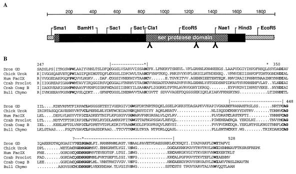
Figure 3.
Alignment of GD with related proteins. (A) Diagram of the gene structure. Region shown in the alignment below is the Ser protease domain with diagonal lines. A hydrophobic secretion signal appears at the N terminus and a hydrophobic tail at the C terminus. Putative N-linked glycosylation sites (NXT/S) are noted by inverted Ys. (B) Each protein is shown beginning with the activating cleavage site (|) and showing the catalytic portion of the protein. Amino acid numbers of the Drosophila protein are shown above the alignment. The putative cleavage site listed for GD is atypical. The H D S residues of the catalytic triad (marked with ∗) are conserved in GD as are most of the Cys bridges with the exception of the penultimate C indicated by (?). A number of other residues are conserved as well. Residues shown in bold correspond to some of the key residues that define distinct structural domains of serine proteases (40). Molecular modeling suggests that the sequence differences seen here could be accommodated into a folded protein that preserved the catalytic site and the substrate groove. The sequences used in this alignment are chicken urokinase P15120, human factor IX clotting enzyme P00740, horseshoe crab proclotting enzyme precursor P21902, crab coagulation factor B A48050, and Bos taurus chymotrypsin National Center for Biotechnology Information sequence ID: 442949.

