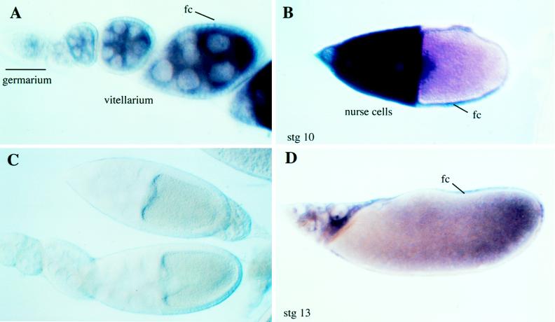
Figure 4.
In situ hybridization to gd mRNA in ovaries. (A) gd mRNA first appears in the germarium. Levels continue to build in the nurse cells and oocytes of the vitellarium. Expression also begins to appear in the follicle cells (fc) as the oocytes move into stage 10. (B) Extensive transcription in both nurse cells and in follicle cells is seen in stage 10 oocytes. In oocytes where the D/V axis can be inferred from the position of the oocyte nucleus, a rough ventral to dorsal gradient of transcript often can be detected. (D) By stage 13, residual gd transcripts remain in the nurse cells as well as the surrounding follicle cells. (C) Sense strand control probe shows no detectable background staining.

