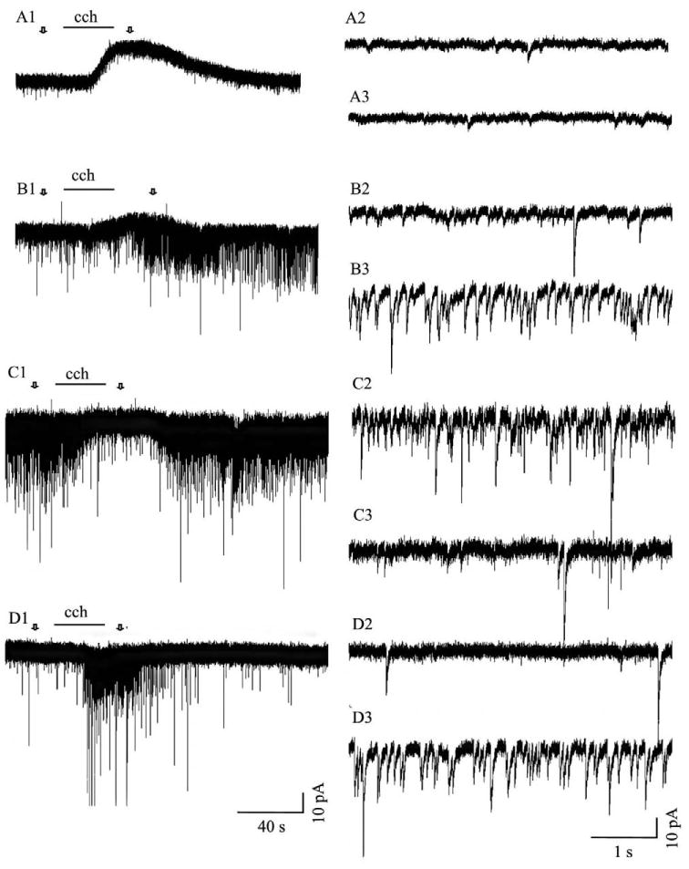
Fig. 3.

Responses in tectal cells evoked by muscarinic receptor activation. Application of carbachol (cch; 100 μM), after desensitization of nicotinic ACL receptors, produced four different responses: an outward membrane current (A1, 57% of the cells), an increase in the frequency and amplitude of sPSCs superimposed upon an outward current (B1, 6% of total recordings), a decrease in the frequency and amplitude of sPSCs (C1, 5% of total recordings) or an increase in the frequency and amplitude of sPSCs (D1, 3% of total recordings). Carbachol did not induce any response in 30% of the cells. Arrows in A1, B1 and C1 indicate areas of traces displayed at a faster time scale prior to drug application (A2, B2 and C2) and during the peak period of the carbachol response (A3, B3 and C3).
