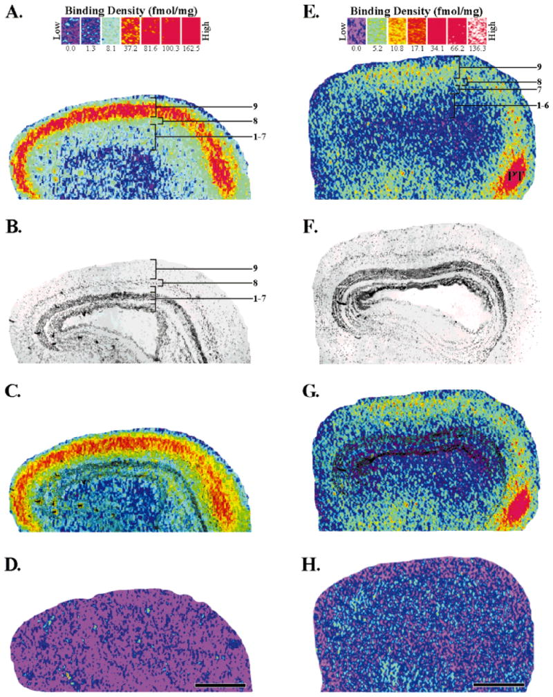
Fig. 2.

Localization of [3H]cytisine (A,C) and [125I]α-bungarotoxin (E,G) binding sites in the adult optic tectum. The binding scales above each column provide a respective index of binding density (fmol/mg wet tissue weight) for each radioligand (see Materials and Methods for details). A: Specific [3H]cytisine (5 nM) binding in the optic tectum. The binding sites were most dense (red) in layer 9. Layer 8 had intermediate densities (yellow/green), whereas layers 1–7 had low densities (light green/light blue). Tectal layers were assigned by using the superimposed image seen in C. B: Brightfield image of the same tissue section that produced the autoradiogram (A). The optic tectum is a laminated structure consisting of alternating cellular and plexiform layers. The darkly stained, main cellular layer is layer 6. C: Superimposition of images A and B which accurately localizes the binding to specific, tectal layers. D: Nonspecific binding, as determined by the addition of nicotine (10 μM) to the incubation solution, was not detected when using [3H]cytisine at 5 nM. E: Total [125I]α-bungarotoxin binding (2.5 nM) in the optic tectum. The densest binding was seen in a pretectal area (PT). Intermediate binding densities were present in tectal layers 8 and 9. F: Brightfield image of the section that produced the autoradiogram in E. G: Superimposition of E and F localizes the binding to the tectal layers. H: Nonspecific [125I]α-bungarotoxin binding as determined by the addition of excess nicotine (10 μM). Anterior is to the right. Scale bars = 500 μm.
