
Fig. 5.
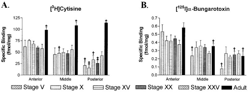
Quantitative analysis of [3H]cytisine and [125I]α-bungarotoxin binding in the retinorecipient layers of the tectum during development. A: Histograms displaying [3H]cytisine (5 nM) binding site densities for anterior, middle, and posterior regions of the optic tectum in tadpoles and adult frogs. Binding densities in every region of the adult tectum were equivalent and significantly higher than those found in any tadpole stage (*P < 0.02). In tadpoles, binding was present at fairly constant levels in the anterior tectum, at slightly lower levels in the middle tectum, and at still lower levels in the posterior tectum. Binding in the posterior tectum tended to increase with development. By stage XXV, binding densities in the posterior tectum were not significantly different from those in the anterior tectum. B: Histograms of [125I]α-bungarotoxin (2.5 nM) binding densities during development. Within the same age group, binding densities were greatest in the anterior tectum, intermediate in middle regions, and lowest in posterior ones. Within each of these regions, binding densities were equivalent across the different age groups. Error bars represent the standard error of the mean (S.E.M.; n = 4). † P < 0.05 compared to the anterior tectum of the same age, and ‡ P < 0.05 compared to both the anterior and middle tectum of the same age.