
Fig. 8.
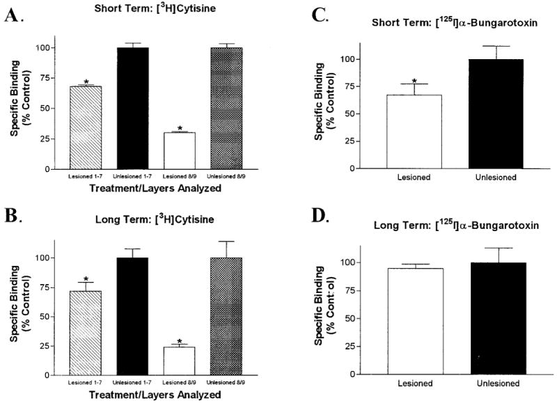
Analysis of [3H]cytisine (5 nM) and [125I]α-bungarotoxin (2.5 nM) binding in the retinorecipient layers of the tectum after lesions of the adult optic nerve. A: Quantitative analysis of [3H]cytisine binding after lesioning of the adult optic nerve with a short survival period. The binding in layers 1–7 of the deafferented lobe was reduced by 31.5 ± 1.5%, whereas the binding in layers 8 and 9 was decreased by 70 ± 1%. B: Long survival periods after optic nerve lesions had a similar effect on [3H]cytisine binding in the deafferented lobe. Binding in layers 1–7 was decreased by 28 ± 8% and that in layers 8 and 9 was reduced by 76 ± 3%. C: Short-term optic nerve lesions had a small, but significant, effect on specific [125I]α-bungarotoxin binding in the superficial layers of the deafferented lobe. Binding was reduced by 33 ± 10% in layers 8 and 9. D: Long-term optic nerve lesions did not affect specific [125I]α-bungarotoxin binding in tectal layers 8 and 9. There was no significant difference between afferented and deafferented lobes. Error bars represent the S.E.M. (*P < 0.05, paired, two-tailed t-test; n = 4 or 5).