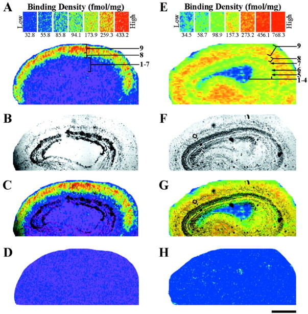
Fig. 7.

Localization of nicotinic (A, C) and muscarinic (E, G) acetylcholine receptor-binding sites in the adult optic tectum. A, Autoradiogram of specific [3H]nicotine (2 nm) binding in the optic tectum. Nicotinic-binding sites are most dense (red–orange) in layer 9. Layer 8 has intermediate densities (yellow–green), whereas layers 1–7 have virtually none (blue–purple). B,Bright-field image of the same tissue section that produced the autoradiogram (A). C,Superimposing A and B with Adobe Photoshop 5.0 accurately localizes the binding to specific, tectal layers. D, Nonspecific binding, as determined by the addition of cytisine (10 μm) to the incubation solution, is not detected when using [3H]nicotine at 2 nm. E, Specific binding of [3H]NMS (2.5 nm) shows the distribution of muscarinic receptors in the optic tectum. The most dense binding (red–orange) is seen in the superficial layers (7–9) and plexiform layer 5, but the binding is less robust (yellow–green) in the layers 1–4 and 6.F, Bright-field image of the section used to produce the autoradiogram in E. G, Layer-specific binding as determined by superimposing E andF. H, Competition of [3H]NMS (2.5 nm) with excess atropine sulfate (10 μm) results in a blank autoradiogram. Scale bar, 500 μm.
