
Figure 4. Impaired BCR-mediated signaling in CD45/CD148 DKO mice.
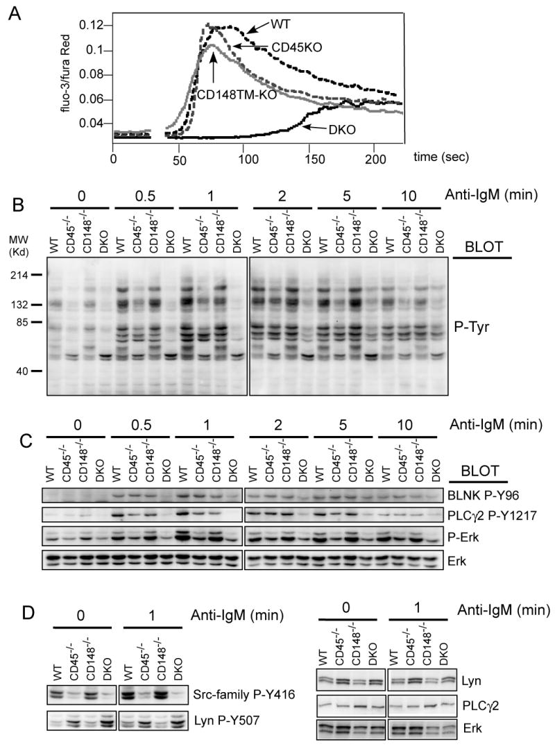
(A) Purified lymph node B cells of the indicated mice were loaded with Fluo3-AM and Fura Red and intracellular free Ca2+ concentrations were monitored before and after addition of IgM F(ab')2 (5μg/ml). (B) Purified lymph node B cells of all four genotypes were stimulated with F(ab')2 fragments of anti-IgM antibody (5 μg/ml). At the indicated time points, the cells were lysed in SDS-PAGE sample buffer and the lysates were analyzed by immunoblotting with phosphotyrosine antibody. (C) Samples from panel (B) were subjected to immunoblotting with phospho-specific antibodies as indicated. Staining of total Erk served as a loading control. (D) Samples from panel (B) (the 0 min and 1 min timepoints) were stained with site-specific antibody against phosphorylated activation loop of SFKs (Src-family P-Y416, this antibody crossreacts with multiple SFKs) and against C-terminal inhibitory phosphotyrosine of Lyn (Lyn P-Y507). The right hand panels are protein controls in the various genotypes. See quantification for westerns shown in (C) and (D) in supplementary Fig. 6.