Figure 3.
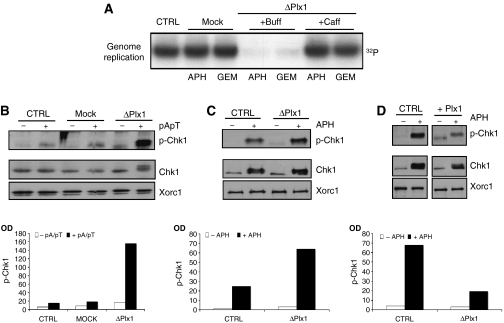
Plx1 suppresses the caffeine-sensitive intra-S-phase checkpoint. (A) The effects of caffeine on DNA replication under stress in the absence of Plx1. Sperm nuclei (7000 nuclei/μl) were replicated in extracts that were untreated (CTRL), mock depleted (Mock) or Plx1 depleted (ΔPlx1) in the presence of 2.5 μM aphidicolin (APH) or 40 nM geminin (GEM). Extracts were supplemented with buffer (Buff) or 5 mM caffeine (Caff). (B) Phosphorylation of Chk1 in untreated (CTRL), mock-depleted (Mock) and Plx1-depleted (ΔPlx1) extracts that were untreated (−) or supplemented with 50 ng/μl pApT (+). Western blot was performed with anti-phospho-Chk1 (p-Chk1), anti-Chk1 and anti-Xorc1 antibodies. Quantification was made by measuring optical density (OD) and is presented as a bar graph. (C) Chk1 phosphorylation in nuclei. Sperm nuclei were incubated with untreated extracts (CTRL) or with Plx1-depleted extracts (ΔPlx1). All extracts were untreated (−) or supplemented with 50 μM aphidicolin (+). The nuclei were isolated and the nuclear proteins were separated by SDS–PAGE. Western blot was performed with anti-phospho-Chk1 (p-Chk1), anti-Chk1 and anti-Xorc1 antibodies. (D) Sperm nuclei were incubated with untreated extracts (CTRL) or with extracts supplemented with 6 ng/μl Plx1 (Plx1) and processed as described above. All extracts were untreated (−) or supplemented with 50 μM aphidicolin (+). As described above, quantification of western blot signals in panels C and D was made by measuring OD and is presented as bar graphs.

