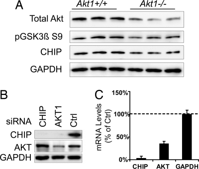
Fig. 3.
Akt1−/− mice have reduced CHIP levels, and decreased Akt levels leads to lower CHIP expression. (A) Brain homogenates from six mice (three Akt1−/− and three Akt+/+) were analyzed by Western blot for total Akt levels, along with CHIP and pGSK3β S9 levels. For each of these proteins, expression was lower in Akt1−/− mice than in Akt1+/+ mice. Quantitation of CHIP reductions by densitometry showed a significant 17% reduction by Student's t test (P = 0.015) (SI Fig. 11A). (B) Hs578T cells transfected with Akt, CHIP, or control siRNAs for 72 h showed reductions in CHIP levels with either CHIP or Akt siRNA. (C) Cells were transfected in triplicate with the same siRNAs described in A. Real-time PCR for CHIP mRNA showed significantly reduced levels (>50%) in cells transfected with Akt siRNA. Primer specificity was confirmed with CHIP siRNA.

