Fig. 6.
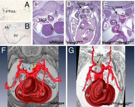
Vangl1gt/+;Vangl2lp/+ embryos exhibit aberrant right subclavian artery. (A) Vangl1 expression in fourth aortic arch of E10.5 Vangl1gt/gt embryo (X-gal staining; magnification, ×600). (B) X-Gal staining of the intracardiac structures of the of E10.5 Vangl1gt/gt embryos (magnification, ×200). (C and D) Transverse sections through the hearts of E14.5 embryos (magnification, ×400). (C) In WT embryos, the right subclavian artery (arrows) is positioned laterally from trachea and esophagus. In Vangl2lp/lp embryos (D) and in Vangl1gt/+;Vangl2lp/+ (E), the right subclavian artery is positioned dorsal to esophagus. (F and G) Three-dimensional reconstruction of the arteries of E14.5 WT (F) and Vangl1gt/+;Vangl2lp/+ hearts (G). Ao, aorta; DAo, dorsal aorta; E, esophagus; LV, left ventricle; PAA, pharyngeal arch artery; PV, pulmonary valve; RA, right atrium; rsca, right subclavian artery; RV, right ventricle; T, trachea; Th, thymic primordium.

