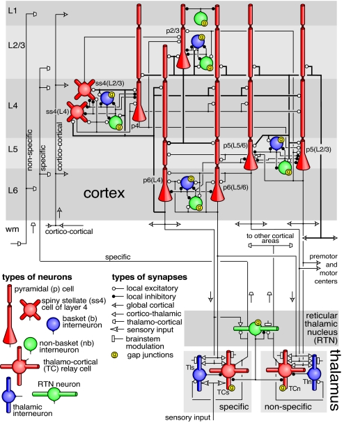
Fig. 2.
Simplified diagram of the microcircuitry of the cortical laminar structure (Upper) and thalamic nuclei (Lower). Neuronal and synaptic types are as indicated. Only major pathways are shows in the figure. Complete details are provided in SI Appendix. L1-L6 are cortical layers; wm refers to white-matter. Arrows indicate types and directions of internal signals.

