
Figure 3.
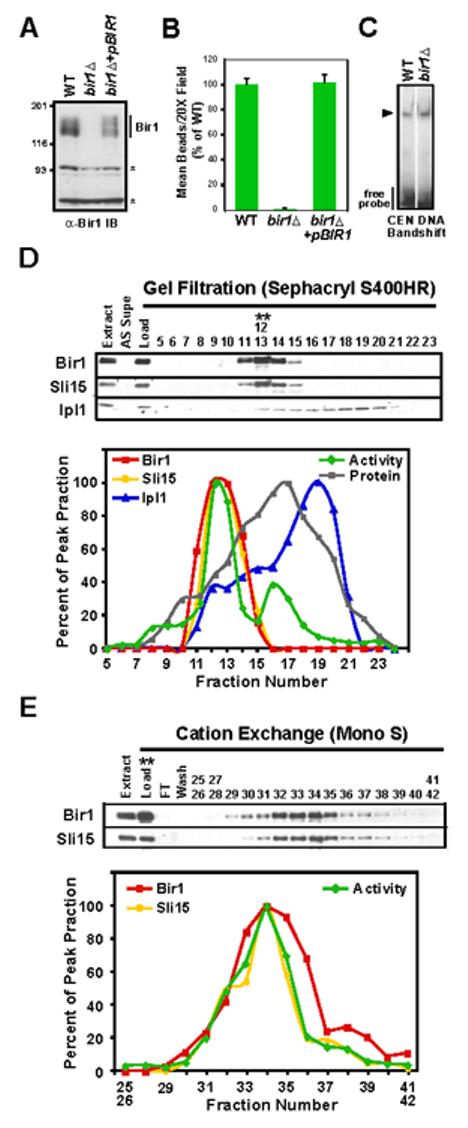
Bir1 is required for linking CEN DNA to microtubules in vitro. (A) bir1∆ cells lack Bir1 protein. Western blot of extracts prepared from WT (ODY49), bir1∆(ODY65), and bir1∆+pCEN-BIR1 (ODY114) strains probed with an anti-Bir1 antibody. Asterisks indicate background bands that serve as loading controls. (B) Bir1 is required for linking CEN DNA to microtubules. Extracts indicated in (A) were analyzed using the bead assay. Activity was normalized relative to the wild-type extract. Error bars=SD. (C) Bir1 does not affect the ability of CBF3 to bind to CEN-DNA. Arrowhead indicates position of the CBF3-CEN DNA complex. (D) Bir1 and Sli15, but not Ipl1, co-fractionate with the complementing activity. Gel filtration fractions of extracts prepared from BIR1:6HA; SLI15:13Myc (ODY97) were analyzed by western blotting using anti-HA, anti-Myc and anti-Ipl1 antibodies. The blot signal intensity for all 3 proteins, as well as activity in the bead assay, is plotted as a percentage of the respective peak fractions (12/13 for Sli15, Bir1, and activity; 18 for Ipl1). (E) Bir1 and Sli15 continue to co-fractionate with the complementing activity during the cation exchange step. The activity peak from gel filtration (fractions 12/13 in (D)) was further fractionated using a MonoS cation exchange column and analyzed as in (D).