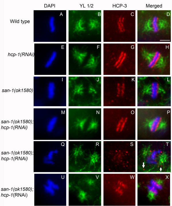
Figure 7.
The san-1(ok1580);hcp-1(RNAi) mitotic blastomeres have normal and abnormal centromere and spindle structure. Analysis of metaphase blastomeres of wild-type (A-D), hcp-1(RNAi) (E-H), san-1(ok1580) (I-L) and san-1(ok1580);hcp-1(RNAi) embryos (M-X) was assayed. Anti-HCP-3 recognizes the centromeric histone HCP-3 and YL1/2 antibody recognizes microtubules. Three typical san-1(ok1580);hcp-1(RNAi) metaphase blastomeres are shown. The metaphase blastomeres shown in Q-X are of abnormal structure. Images were collected using a spinning disc confocal microscope. Arrow points to HCP-3 not associated with the chromosomes. Bar = 2 μm.

