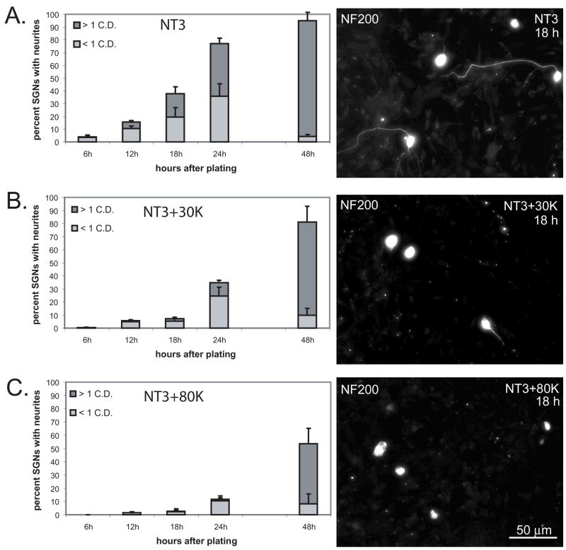
Figure 2.
Membrane depolarization delays SGN neurite initiation. Spiral ganglion cultures were maintained in NT-3 (A), NT-3+30K (B), or NT-3+80K (C) for 6, 12, 18, 24 and 48 hr after plating, fixed, and immunolabeled with anti-NF-200. The percentage of SGNs bearing a neurite < 1 cell diameter (C.D.) or > 1 C.D. was determined for each timepoint. The data presented represent the average of the 3 repetitions and error bars present standard deviation. At 12 hr, a significantly higher percentage of SGNs in NT-3 had a neurite compared with those in NT-3+30K (p<0.001, ANOVA followed by post-hoc Holm-Sidak methodand those in NT-3+80K (p<0.001). Similarly, at 12 hr a significantly higher percentage of SGNs in NT-3+30K had a neurite compared with those in NT-3+80K (p<0.001). These differences persisted until the 48 hr time point at which point there was no longer a statistically significant difference between the percent of SGNs bearing a neurite in NT-3 compared with those in NT-3+30K (p=0.057). Right panels present representative images of cultures immunostained with anti-NF-200 and maintained in NT-3 (A), NT-3+30K (B), and NT-3+80K (C) for 18 h. Scale bar=50 μm.

