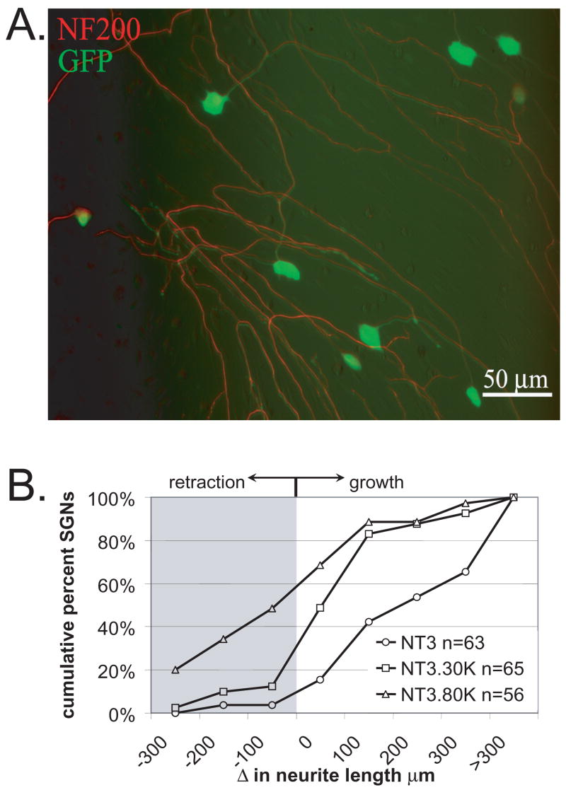
Figure 3.
Membrane depolarization inhibits SGN neurite extension. Spiral ganglion cultures were maintained in NT-3 (50 ng/ml) for 48 hr to allow for neurites to develop. The cultures were then treated with a feline immunodeficiency lentiviral vector encoding green fluorescent protein (FIV-GFP). The GFP fills the entire soma and neurite processes. Initial images of SGNs were captured based on GFP fluorescence. The cultures were then maintained in NT-3, NT-3+30K, or NT-3+80K for an additional 24 h, fixed, labeled with anti-NF-200 to confirm that imaged processes represented SGN neurites, and reimaged. A. Spiral ganglion culture treated with FIV-GFP (green) and labeled with anti-NF-200 followed by an Alexa 568 secondary antibody (red) demonstrating that FIV-GFP transduces a high percentage of SGNs and that the GFP fills the entire neurite process. In this composite image, the green and red images were offset slightly to better illustrate the overlap of GFP and NF-200 labeling. Scale bar=50 μm. B. Change in neurite length over a 24 hr interval for cultures maintained in NT-3, NT-3+30K, or NT-3+80K presented as cumulative percent histograms. Values <0 μm represent neurite retraction while values >0 μm represent neurite growth. Each condition was repeated three times and is significantly different (p<0.05) than the others by Kruskal-Wallis ANOVA on Ranks followed by a Dunn’s post-hoc comparison. n=cumulative number of SGNs scored for each condition.

